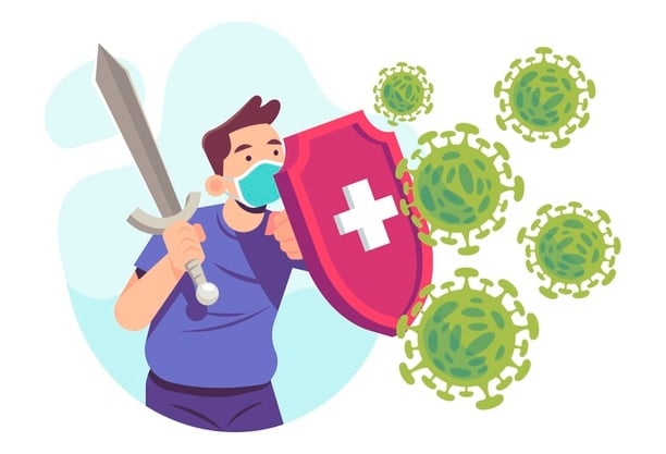

THE WARFARE: PART 2 THE FIGHT BETWEEN THE VIRUS AND THE HOST
Harish Kumar Senapati,
Prakhar Varshney,
Prikshit,
Prachiti Vithole,
Pranav Pradhan
INTRODUCTION
So far… SARS-COV-2 binds to the host cell ACE2 receptor by means of its spike glycoprotein (S protein) to enter the cytoplasm where it releases its RNA ssRNA genome and initiates various processes leading to its replication and pathogenesis [Refer to the ‘first article of Warfare series of CONOCIMIENTO, ISSUE 2’ for more details].
In the second part of ‘The Warfare’ series, we have summarized some important findings from recent literature on host’s immune response against viral pathogenesis.
PATHOGENESIS AND CORRESPOND INGIMMUNE RESPONSE : AN OVERVIEW
The viral antigen is presented by antigenpresenting cells (APCs) with the aid of major histocompatibility complex (MHC). Antigen presentation stimulates both cellular and humoral immunity [Figure 1].
Immune effector cells release large amounts of cytokines (protein molecule that mediates and regulate inflammation) & chemokines (cheimcals that attract other cells) that might rapidly induce acute respiratory distress syndrome (ARDS), single or multiple organ failure & eventually death.
The immune system is a complex network of cells and proteins that defends the body against infection. It keeps a record of every microbe it has ever encountered, so that it can recognize and destroy the pathogen quickly if it enters the body again. Our body inherently possesses innate immunity and can also subsequently develop adaptive immunity.
Innate immune cells, e.g., macrophages, mast cells attack the pathogen as soon as it enters the body. Adaptive immune cells (B & T lymphocytes) are activated by the APCs, that promotes production of chemokines and cytokines including IFN (interferons), IP (induced protein), MCP (monocyte chemo attractant protein), IL (interleukins) etc. Refer to the textbook ‘IMMUNOLOGY’ by Barbara A. Osborne & Janis Kuby for a better understanding.
PERSPECTIVES ON IMMUNE RESPONSES
SARS-CoV-2 infection can be roughly divided into three stages:
Stage I, an asymptomatic incubation period with or without detectable symptoms;
Stage II, non-severe symptomatic period indicating the presence of virus;
Stage III, severe respiratory symptomatic stage with high viral load.
One of the biggest unanswered questions is why some individuals develop severe disease while others do not.
Although the early immune response is positive and leads to viral clearance, the virus being a novel one, innate immunity cannot efficiently eradicate it from the body and gets the vulnerable tissues infected (See Warfare Series Part 1). During the incubation and non-severe stages, adaptive immune response is activated to eliminate the virus and to stop disease progression to severe stages. For the development of an endogenous protective immune response, the host should be in good general health and an appropriate genetic background, that elicits specific antiviral immunity. However, when protective immune response is impaired, the virus will propagate and massive destruction of the affected tissues will occur, especially in organs such as lungs, intestine and kidney. The damaged cells induce inflammation in the lungs by macrophages and granulocytes. Lung inflammation is the main cause of life-threatening respiratory disorders at the severe stage. The exaggerated inflammatory response is the result of cytokine storm (abnormal production of cytokine & chemokines) caused by immune dysregulation.
A study by Chen et al. (2020) on virus dynamics and host response included a report on the viral load and antibody profiles of a cohort of 23 COVID-19 patients. The viral load peaked during the first week of illness; then, gradually declined over the second week. Viral load was also shown to correlate with age. Furthermore, both IgG & IgM antibodies started to increase around day 10 after symptoms onset and most patients had seroconversion within the first 3 weeks. Finally, the IgG & IgM antibodies levelled against the SARS-CoV-2 nucleoprotein.
The high viral load in elderly patients is associated not only with low immunity, but also with high expression of the ACE2 receptor in adults. The most common symptoms at onset of COVID-19 illness are fever, cough & fatigue, while other symptoms include sputum production, headache, hemoptysis, diarrhea, dyspnea & lymphopenia. Clinical features revealed by a chest CT scan is examined to be similar to pneumonia. [Refer to COVID 19 Etiology article in CONOCIMIENTO 1’ for more details] However, other abnormalities such as RNAaemia (presence of RNA in the serum), acute respiratory distress syndrome and incidence of ground-glass opacities (the quality or state of a body that makes it impervious to the rays of light) that led to death, were observed as well.
Histopathological observations of pulmonary lesions in SARS cases not only showed nonspecific inflammatory responses such as edema and cell infiltration, but also, severe exfoliation of alveolar epithelial cells, alveolar septal widening, damage to alveolar septa & alveolar space infiltration in a distinctly organized manner were found. Pathologically, inflammation includes degeneration (necrosis), infiltration, and hyperplasia.
CONCLUSION
Research on Coronavirus is only about two decades old, after the emergence of SARS and MERS. Although most studies on the virus is currently focused on therapeutics, vaccines, other epidemiological aspects also need to be considered. Some studies claim that there are thousands of other coronaviruses currently circulating in bats, indicating possible future spill over events and outbreaks. Hence, more research on basic and applied virology of coronaviruses is the need of the hour to prevent the current pandemic as well as to be ready for ‘the warfare’ in long run.
REFERENCES
1. Chen, Y., & Li, L. (2020). SARS-CoV-2: virus dynamics and host response. The Lancet Infectious Diseases, 20(5), 515-516.
2. Katze, M. G., Korth, M. J., Law, G. L., & Nathanson, N. (Eds.). (2015). Viral Pathogenesis: From Basics to Systems Biology. Academic Press.
3. Li, G., Fan, Y., Lai, Y., Han, T., Li, Z., Zhou, P., ... & Zhang, Q. (2020). Coronavirus infections and immune responses. Journal of medical virology, 92(4), 424-432.
4. Lin, L., Lu, L., Cao, W., & Li, T. (2020). Hypothesis for potential pathogenesis of SARS-CoV-2 infection–a review of immune changes in patients with viral pneumonia. Emerging microbes & infections, 9(1), 727-732.
5. Li, X., Geng, M., Peng, Y., Meng, L., & Lu, S. (2020). Molecular immune pathogenesis and diagnosis of COVID-19. Journal of Pharmaceutical Analysis.
6. Rothan, H. A., & Byrareddy, S. N. (2020). The epidemiology and pathogenesis of coronavirus disease (COVID-19) outbreak. Journal of autoimmunity, 102433.
7. Sarzi-Puttini, P., Giorgi, V., Sirotti, S., Marotto, D., Ardizzone, S., Rizzardini, G., ... & Galli, M. (2020). COVID-19, cytokines and immunosuppression: what can we learn from severe acute respiratory syndrome?. Clinical and experimental rheumatology, 38(2), 337-342.
8. Shi, Y., Wang, Y., Shao, C., Huang, J., Gan, J., Huang, X., ... & Melino, G. (2020). COVID-19 infection: the perspectives on immune responses.
WEBSITES REFERRED
https://www.youtube.com/watch?v=8_bOhZd6ieM
https://www.vectorstock.com/royalty-free-vector/young-manwith-sword-fight-with-coronavirus-vector-29340043 https://app.biorender.com/biorender-templates https://www.theweek.in/theweek/cover/2020/03/19/whenthe-wild-moves-in.html https://onlinelearning.hms.harvard.edu/hmx/immunity/
Website Designed By- Tech Team Episteme
©️Episteme Science Magazine 2022